Visualizing Tissues and Gene Products
Ab Staining
Acetylated Tubin Cilia Staining: X. tropicalis larvae: This protocol works well for epidermal cilia on Xenopus embryos. It was adapted from a protocol provided by Basudha Basu.
General Immuno Staining Protocol: Lists primary and secondary antibodies for muscle, mitotic and lymphatic cells, and notochord.
Cartilage Dissection
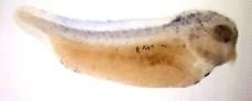
Cartilage Dissection: Once cartilage has been stained, dissection of the facial cartilage allows for efficient visualization and subsequent analysis of cranial cartilage formation.
Cartilage Staining
This protocol is adapted from one kindly provided to us by Dr. Carol Labonne
Cartilage Staining: To use a basic water-based dye to stain for acid mucosubstances and acetic mucins located on the cartilage of fixed embryos, permitting the examination of cartilage formation.
TUNEL detects cells undergoing apoptosis. Please see the paper by Hensey and Gautier for the details. In our hands, we detect very few if any apoptotic cells during normal development in Xenopus tropicalis using this protocol. However, the protocol is effective in identifying apoptotic cells in embryos that are undergoing substantial apoptosis.
In Situ Hybridization
The X. laevis procedure for whole mount in situ hybridization works perfectly well for X. tropicalis embryos without change. We use the protocol by Sive et al. Early Development of Xenopus laevis: A Laboratory Manual CSHL Press (2000).
In order to efficiently perform the procedure, we routinely place embryos in porous baskets to facilitate the solution exchanges. Links to the detailed and short protocols, as well as links for making RNA probe:
Protocols for In Situ Hybridization:
Whole Mount In Situ Hybridization Protocol
Whole Mount In Situ Hybridization - Short Worksheet
Whole Mount In Situ Hybridization - Short and Fast Protocol
Protocol for Making Antisense RNA probe (dig labelled):
RNA Probe Synthesis for In Situ Hybridizations - Condensed Protocol
Standard Protocol Modifications
Eliminate PK
In an attempt to streamline the procedure, we have compared the original protocol to a modified protocol with particular steps omitted. In a side by side comparison, omission of the proteinase K step led to a significant reduction in signal especially in neural and endodermal staining.
Eliminate Pre-hybridization
The first day of the in situ protocol is considerably prolonged because of the long pre-hybridization step (six hours). We have tested the minimum length of time required for pre-hybridization. In X. laevis embryos, the prehybridization step may be reduced from six to four hours but must last for at least four hours. Shorter pre-hyb times led to a degradation in signal.
Using a subset of probes we have found that in X. tropicalis embryos the pre-hybridization step appears unnecessary and equivalent signal can be obtained if the pre-hybridization is omitted, reduced to one hour, or kept at the full six hours. Eliminating the pre-hybridization step greatly shortens the first day of the protocol. Of course only a limited number of probes were tried, and we recommend comparing the standard procedure with and without pre-hybridization to be sure that the signal is equivalent. In routine practice, we pre-hyb for one hour at a minimum which works well and is convenient.